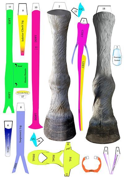
Anatomy of the Equine Distal Limb
This is a detailed paper chart displaying the anatomy of the distal limb of a horse from both a dorsal and palmar view. It illustrates the relationship between the common and lateral digital extensor tendons and their attachment to the bones, as well as the relation between the superficial and deep digital flexor tendons. Other structures such as the inferior check ligament, the manica flexoria, the palmar annular ligament, proximal and distal annular ligaments, and the collateral cartilages are also demonstrated. Furthermore, the chart arranges the sesamoidean ligaments into three layers, showing straight, oblique, and cruciate sesamoidean ligaments. The deepest layer provides a view of the navicular bursa, navicular bone, other bony structures, and the distal limb joints. This chart is a vital tool for anyone who works with horses as it provides a clear understanding of the topographical anatomy of the distal limb, which is essential for interpreting imaging modalities such as radiography and ultrasonography. It also helps horse owners and professionals to identify the location of certain pathological cases, such as navicular disease.
